Scrotal calcinosis is a rare, benign condition characterized by the presence of multiple calcified nodules within the skin of the scrotum. Though generally harmless, the condition can cause discomfort and cosmetic concerns, prompting patients to seek medical attention. This article provides an in-depth look at scrotal calcinosis, including its causes, symptoms, diagnosis, and treatment, with a particular focus on surgical intervention.
What is Scrotal Calcinosis?
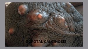
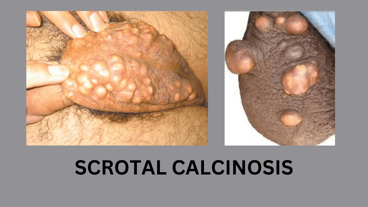
Scrotal calcinosis is a dermatological condition in which calcium deposits, or calcified nodules, develop in the scrotal skin. These nodules are typically asymptomatic, but they can sometimes cause irritation, pain, or discomfort, particularly if they become infected or ulcerate. The nodules may vary in size and number, and their growth is generally slow.
The condition primarily affects men in their third or fourth decade of life but can occur at any age. While the exact cause remains unclear, scrotal calcinosis is often considered a form of idiopathic calcinosis, meaning the reason for the calcium deposits is unknown.
Causes of Scrotal Calcinosis
There is ongoing debate about the underlying cause of scrotal calcinosis. Several theories have been proposed:
- Idiopathic Calcinosis: In many cases, no underlying cause can be identified, and the nodules develop without any preceding event or condition. This is referred to as idiopathic scrotal calcinosis.
- Degeneration of Epidermal Cysts: Some researchers suggest that scrotal calcinosis may result from the degeneration of pre-existing epidermal cysts. These cysts, which form within the skin, may rupture and undergo calcification over time.
- Metabolic Disorders: Certain metabolic conditions, such as hypercalcemia (high calcium levels in the blood), may contribute to the development of calcinosis. However, this is rare in cases of scrotal calcinosis.
- Infections and Inflammation: Chronic infections or inflammation in the scrotal area may lead to calcium deposition as a response to tissue damage.
- Genetic Factors: Some studies have pointed to the possibility of a genetic predisposition to scrotal calcinosis, though this has not been conclusively proven.

Symptoms of Scrotal Calcinosis
Scrotal calcinosis often presents with the following signs and symptoms:
- Nodules or Lumps: The primary symptom is the presence of firm, painless nodules on the scrotum. These nodules may vary in size from a few millimeters to several centimeters.
- Discomfort or Pain: In some cases, especially when the nodules are large or numerous, the patient may experience discomfort or pain in the scrotal area. This can be exacerbated by friction or physical activity.
- Ulceration: In rare cases, the nodules may become ulcerated, leading to infection or discharge from the affected area.
- Cosmetic Concerns: While scrotal calcinosis is usually harmless, the appearance of the nodules can cause distress for patients due to their unsightly nature.
- Itching: Sometime itching may be predominant feature. Patient visited to clinic with itching only.
Diagnosis of Scrotal Calcinosis
The diagnosis of scrotal calcinosis is typically made through clinical examination and patient history. The doctor will examine the scrotum for the presence of calcified nodules and may ask about the duration of symptoms and any potential underlying conditions.
To confirm the diagnosis and rule out other conditions, the following tests may be conducted:
- Biopsy: A small sample of the affected skin may be removed and analyzed under a microscope to confirm the presence of calcium deposits and to rule out other skin conditions or tumors.
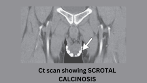
- Imaging: In some cases, imaging tests such as ultrasound or X-rays may be used to visualize the extent of the calcification and to determine the size and number of nodules.
- Blood Tests: Blood tests may be conducted to assess calcium levels and to rule out any metabolic disorders that could be contributing to the calcification.

Surgical Treatment of Scrotal Calcinosis
While scrotal calcinosis is generally benign and does not require treatment unless it causes discomfort or cosmetic concerns, surgical intervention is the most effective option for removing the calcified nodules. Surgery is typically recommended for patients who experience pain, recurrent infections, or significant distress due to the appearance of the nodules.
Surgical Procedure
The surgical treatment of scrotal calcinosis involves the excision (removal) of the calcified nodules. The procedure is relatively simple and is usually performed under local anesthesia. Here’s an overview of the steps involved:
- Anesthesia: Local anesthesia is administered to numb the scrotal area, ensuring that the patient does not feel pain during the procedure.
- Incision and Removal: The surgeon makes small incisions in the scrotal skin to access the calcified nodules. The nodules are carefully removed, ensuring that all calcified material is excised to prevent recurrence.
- Closure: After the nodules are removed, the incisions are sutured (stitched) closed. In some cases, dissolvable stitches may be used, while in others, the patient may need to return to have the stitches removed.
- Postoperative Care: After the procedure, the patient is advised to rest and avoid physical activity that could cause trauma to the scrotal area. The surgeon may prescribe antibiotics to prevent infection, and pain relievers may be recommended to manage postoperative discomfort.
Recovery
Recovery from surgery for scrotal calcinosis is typically straightforward. Most patients can resume normal activities within a week, though heavy lifting and strenuous exercise should be avoided for a few weeks to allow the area to heal.
Patients are advised to monitor the surgical site for signs of infection, such as redness, swelling, or discharge. If any of these symptoms occur, they should contact their doctor for further evaluation.
Complications and Risks of Surgery
While surgery for scrotal calcinosis is generally safe, there are some potential risks and complications, including:
- Infection: As with any surgical procedure, there is a risk of infection at the incision site. Proper wound care and antibiotics can help minimize this risk.
- Scarring: Scarring is a possibility, though the scars are usually small and fade over time.
- Recurrence: In rare cases, calcified nodules may recur after surgery, especially if any residual calcified material is left behind during the procedure.
Alternatives to Surgery
In cases where the nodules are asymptomatic and do not cause discomfort or cosmetic concerns, patients may choose to forgo surgery. Regular monitoring and good hygiene practices can help prevent complications such as infection or ulceration.
However, there are no effective non-surgical treatments for scrotal calcinosis. Medications, topical creams, and other conservative treatments are generally ineffective in managing the condition.
Conclusion
Scrotal calcinosis is a rare but benign condition characterized by the presence of calcified nodules in the scrotal skin. While it typically does not cause significant symptoms, some patients may experience discomfort, pain, or cosmetic concerns. Surgical excision is the most effective treatment for removing the nodules and providing relief.
If you suspect you have scrotal calcinosis or are concerned about lumps on your scrotum, it is essential to consult a healthcare provider for proper diagnosis and management. While the condition is generally harmless, prompt treatment can alleviate symptoms and prevent potential complications.

